| info |
| WORK |
| photos |
Current interests
GLSL (OpenGL), OpenCL, multi-threaded (CPU) programming, clusters, and their applications to medical imaging and image processing. Focusing especially in the topics of data analysis, image registration, resampling, segmentation, surface modelling, atlases, DTI, and visualization.
Sample images from Voxlab (work in progress)
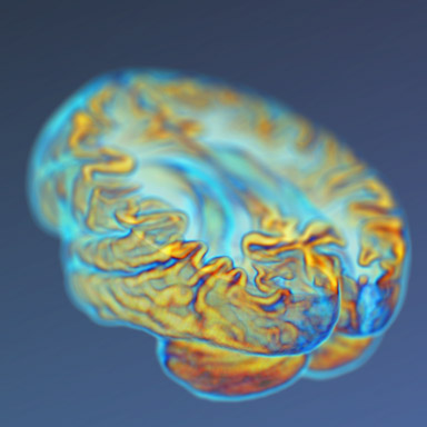
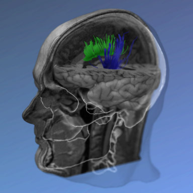
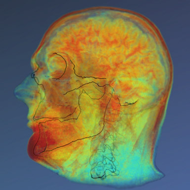
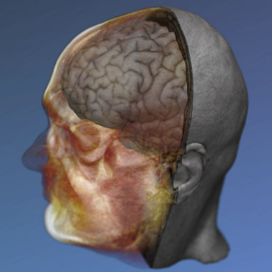
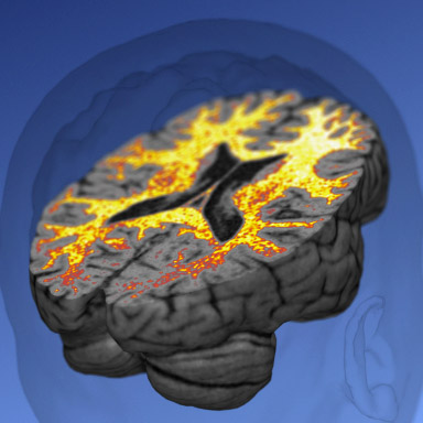

See more images in Voxlab pages.
Articles in international peer-reviewed journals
- Boldt R., Malinen S., Seppä M., Tikka P., Savolainen P., Hari R., Carlson S. Listening to an Audio Drama Activates Two Processing Networks, One for All Sounds, Another Exclusively for Speech. PloS ONE, 8(5): e64489, 2013.
- Pamilo S., Malinen S., Hlushchuk Y., Seppä M., Tikka P., Hari R. Functional Subdivision of Group-ICA Results of fMRI Data Collected during Cinema Viewing. PloS ONE, 7(7): e42000, 2012.
- Hiltunen J., Seppä M., Hari R. Evaluation of voxel-based group-level analysis of diffusion tensor images using simulated brain lesions. Neuroscience Research, 71, 377-386, 2011.
- Seppä M. Continuous sampling in mutual-information registration. IEEE Transactions on Image Processing, 17, 823-826, 2008.
- Seppä M. High-quality two-stage resampling for 3-D volumes in medical imaging. Medical Image Analysis, 11, 346-360, 2007. (C code)
- Korvenoja A., Kirveskari E., Aronen H.J., Avikainen S., Brander A., Huttunen J., Ilmoniemi R.J., Jääskeläinen J.E., Kovala T., Mäkelä J.P., Salli E., Seppä M. Sensorimotor cortex localization: Comparison of magnetoencephalography, functional MR imaging, and intraoperative cortical mapping. Radiology, 241, 213-222, 2006.
- Hiltunen J., Suortti T., Arvela S., Seppä M., Joensuu R., Hari R. Diffusion tensor imaging and tractography of distal peripheral nerves at 3 T. Clinical Neurophysiology, 116, 2315-2323, 2005.
- Seppä M., Hämäläinen M. Visualizing human brain surface from T1-weighted MR images using texture-mapped triangle meshes. NeuroImage, 26, 1-12, 2005.
- Forss N., Raij T., Seppä M., Hari R. Common cortical network for first and second pain. NeuroImage, 24, 132-142, 2005.
- Tarkiainen A., Liljeström M., Seppä M., Salmelin R. The 3D topography of MEG source localization accuracy: Effects of conductor model and noise. Clinical Neurophysiology, 114, 1977-1992, 2003.
- Mäkelä J., Kirveskari E., Seppä M., Hämäläinen M., Forss N., Avikainen S., Salonen O., Salenius S., Kovala T., Randell T., Jääskeläinen J., Hari R. Three-Dimensional Integration of Brain Anatomy and Function to Facilitate Intraoperative Navigation Around the Sensorimotor Strip. Human Brain Mapping, 12, 180-192, 2001.
- Vanni S., Tanskanen T., Seppä M., Uutela K., Hari R. Coinciding early activation of the human primary visual cortex and anteromedial cuneus. Proceesings of the National Academy of Sciences of the United States of America, 98, 2776-2780, 2001.
- Hari R., Hänninen R., Mäkinen T., Jousmäki V., Forss N., Seppä M., Salonen O. Three hands: fragmentation of human bodily awareness. Neuroscience Letters, 240, 131-134, 1998.
- Uusitalo M., Williamson S., Seppä M. Dynamical organisation of the human visual system revealed by lifetimes of activation traces. Neuroscience Letters, 213, 149-152, 1996.
Images employ FancyBox library and some of the visualization use SRI24 Atlas data.